Proximity Ligation Assay: A breakthrough in cell biology research

Cellular processes can only be understood as the dynamic interplay of proteins. This means that we need accurate techniques to monitor interactions of endogenous proteins directly in individual cells and tissues, to reveal the cellular and molecular architecture and its responses to disturbances. 

Proximity Ligation Assay is a cutting-edge technology that has revolutionized the way researchers study protein interactions and protein interplay. 

In traditional methods, detecting protein interactions and cellular localization involved laborious and time-consuming procedures such as immunoprecipitation, BRET, and cross-linking methods. The advent of Proximity Ligation Assay has made it possible to perform these experiments in a faster and more efficient manner, with high sensitivity and specificity. 

Proximity Ligation Assay principle

The principle behind proximity ligation assay is based on a combination of immunoassay and DNA amplification. When antibodies conjugated to oligonucleotides (called Navenibodies) bind protein targets in close proximity, their conjugated oligonucleotides can template circularization of an added pair of oligonucleotides via enzymatic ligation. The circularized DNA strands remain hybridized to the Navenibodies, and one of the oligonucleotides serves as a primer for a localized rolling circle amplification (RCA) reaction. The resulting amplified DNA is detected as a bright fluorescent or chromogenic dot by suitably labeled detection probes.  

One of the key advantages of proximity ligation assay is that it allows for the study of protein interactions in situ, without the need for protein purification or separation. This results in a more accurate representation of the protein interactions that occur in cells and tissues. 

The use of proximity ligation assay has grown rapidly in recent years, with applications in a wide range of areas such as cancer research, neuroscience, and developmental biology. Some of the popular applications of proximity ligation assay include the study of protein-protein interactions, the determination of subcellular localization, and the analysis of protein modification. 

In situ proximity ligation has been applied to improve sensitivity, specificity, and target range in many methods for localized protein detection besides for microscopy, for example, western blotting, flow cytometry and sandwich enzyme-linked immunosorbent assays (ELISA). This technology is positioned to play an integral part in spatial biology research and clinical applications, for high-performance protein detection in situ, and to study protein-protein interactions and post-translational modifications directly in the intact tissue microenvironment. 

Naveni Technology the next-generation Proximity Ligation Assay

In 2018, Axel Klaesson et al., published a paper describing a new proximity probe design called UnFold, based on new, patented oligonucleotide sequences. The UnFold technique differs from the first generation in situ PLA (Duolink® Sigma) technique in that all DNA elements required for amplified detection are included in the oligonucleotides conjugated to the antibodies.  

Compared to the conventional in situ PLA probe design, the UnFold in situ proximity probes improve the efficiency of signal generation: The oligonucleotide that can form a circle is already present on one of the probes, and only one ligation reaction is required, which increases the likelihood that circles can form that are capable of serving as template for RCA. The UnFold technique is commercialized under the trademark Naveni™Flex by Navinci, which is the new name of Olink Biosciences. 

Proximity Ligation Assay principle

Primary antibody incubation:  Two primary antibodies bind to their target epitopes, located on a single protein or two nearby proteins. Navenibody incubation: Navenibodies (carefully selected antibodies conjugated to proprietary oligonucleotide arms) bind their respective primary antibodies. Circle formation: Only if the Navenibodies are in close proximity the attached oligos can generate a DNA circle guaranteeing high specificity by reducing nonspecific background staining. Using the proprietary NaveniTM technology, the efficacy of the oligo design improves the signal strength, ensuring high sensitivity. Amplification and detection: By the addition of polymerase, a rolling circle amplification process is initiated. Fluorescent or HRP/AP labeled probes are bound to the amplified DNA, generating a fluorescence or a chromogenic dot after adding substrate. The high signal-to-noise enables the detection of separate proximity events, allowing for a resolution down to a single protein or protein-protein interaction.

Proximity Ligation Assay principle

Primary antibody incubation

Two primary antibodies bind to their target epitopes, located on a single protein or two nearby proteins.

Navenibody incubation

Navenibodies (carefully selected antibodies conjugated to proprietary oligonucleotide arms) bind their respective primary antibodies.

Circle formation

Only if the Navenibodies are in close proximity the attached oligos can generate a DNA circle guaranteeing high specificity by reducing nonspecific background staining. Using the proprietary NaveniTM technology, the efficacy of the oligo design improves the signal strength, ensuring high sensitivity.

Amplification and detection

By the addition of polymerase, a rolling circle amplification process is initiated. Fluorescent or HRP/AP labeled probes are bound to the amplified DNA, generating a fluorescence or a chromogenic dot after adding substrate. The high signal-to-noise enables the detection of separate proximity events, allowing for a resolution down to a single protein or protein-protein interaction.

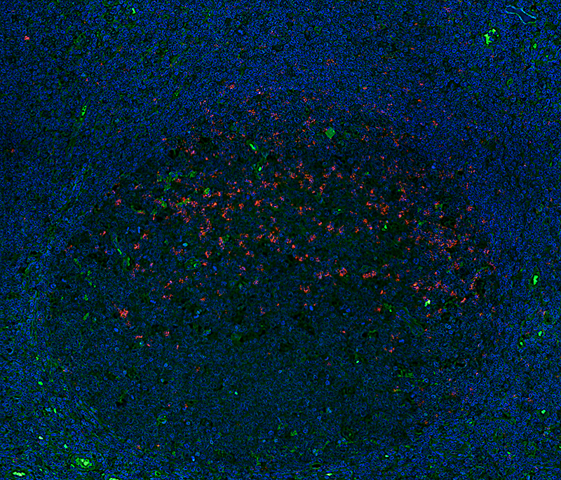
PD1/PD-L1 interaction detected with NaveniFlex Tissue

Innovation ongoing

Proximity Ligation Assay principle

Navinci is a strong research organization and as the center of excellence for proximity ligation technology, we continue to innovate the technology with new developments and improvements. A number of additional patent applications have been added, and we now have a broad product portfolio based on our Naveni Proximity Ligation Technology. See all products here. 

Examples of the latest innovations are several. Naveni™TriFlex, a revolutionary proximity-based technology that concurrently visualizes two proteins in a free and interacting within any cell compartment. The detected protein A, B and the interaction of A/B signals are amplified and generate fluorescent readout in three different channels corresponding to each protein pool. The TriFlex method gives more information from the same experiments and will make it possible to unravel e.x. protein degradation and protein interplay. Learn more about Naveni™TriFlex here. 

With a proprietary solution, we have also solved a difficult problem in the immunofluorescent staining of tissues. Tissues are complex multicellular structures where fluorescently labeled detection reagents have been observed to bind unspecifically to certain cell structures resulting in background staining that can be difficult to distinguish from true biologically relevant staining. To address this problem, we developed a new proprietary version of NaveniFlex, highly optimized for tissue use. The product line tailor-made for tissue is called NaveniFlex Tissue. 

We are experts in assay development and as we know that it takes time to find the optimal primary antibodies. We have expanded our product line of target-specific kits with primary antibodies included. Our latest focus is on target-specific kits within immunooncology, we have a kit for the detection of PD1/PD-L1 interaction, designed to help researchers better understand the interactions between immune checkpoint proteins and cancer cells and how these interactions play a role in the development and treatment of cancer. Read more about PD1 signaling. 

Phosphorylation of proteins are important part of the signaling cascades, but studying phosphorylations has been difficult due to the unspecificity of phospho antibodies. The target-specific Naveni PTMs utilized the specificity with Proximity Ligation Assay and includes validated primary antibodies to detect phosphorylated proteins such as PD1, VEGFR2, HER2 etc. To Target specific kits. 

In conclusion, the proximity ligation assay has become an indispensable tool in biomedical research, providing researchers with a fast, efficient, and sensitive method to study protein interactions and protein localization on single-cell level. With innovations like the Naveni technology leading the way in the development and commercialization of proximity ligation assay technology, the future of biomedical research looks bright. 

Innovation ongoing

Navinci is a strong research organization and as the center of excellence for proximity ligation technology, we continue to innovate the technology with new developments and improvements. A number of additional patent applications have been added, and we now have a broad product portfolio based on our Naveni Proximity Ligation Technology. See all products here. 

Examples of the latest innovations are several. Naveni™TriFlex, a revolutionary proximity-based technology that concurrently visualizes two proteins in a free and interacting within any cell compartment. The detected protein A, B and the interaction of A/B signals are amplified and generate fluorescent readout in three different channels corresponding to each protein pool. The TriFlex method gives more information from the same experiments and will make it possible to unravel e.x. protein degradation and protein interplay. Learn more about Naveni™TriFlex here. 

With a proprietary solution, we have also solved a difficult problem in the immunofluorescent staining of tissues. Tissues are complex multicellular structures where fluorescently labeled detection reagents have been observed to bind unspecifically to certain cell structures resulting in background staining that can be difficult to distinguish from true biologically relevant staining. To address this problem, we developed a new proprietary version of NaveniFlex, highly optimized for tissue use. The product line tailor-made for tissue is called NaveniFlex Tissue. 

We are experts in assay development and as we know that it takes time to find the optimal primary antibodies. We have expanded our product line of target-specific kits with primary antibodies included. Our latest focus is on target-specific kits within immunooncology, we have a kit for the detection of PD1/PD-L1 interaction, designed to help researchers better understand the interactions between immune checkpoint proteins and cancer cells and how these interactions play a role in the development and treatment of cancer. Read more about PD1 signaling. 

Phosphorylation of proteins are important part of the signaling cascades, but studying phosphorylations has been difficult due to the unspecificity of phospho antibodies. The target-specific Naveni PTMs utilized the specificity with Proximity Ligation Assay and includes validated primary antibodies to detect phosphorylated proteins such as PD1, VEGFR2, HER2 etc. To Target specific kits. 

In conclusion, the proximity ligation assay has become an indispensable tool in biomedical research, providing researchers with a fast, efficient, and sensitive method to study protein interactions and protein localization on single-cell level. With innovations like the Naveni technology leading the way in the development and commercialization of proximity ligation assay technology, the future of biomedical research looks bright. 
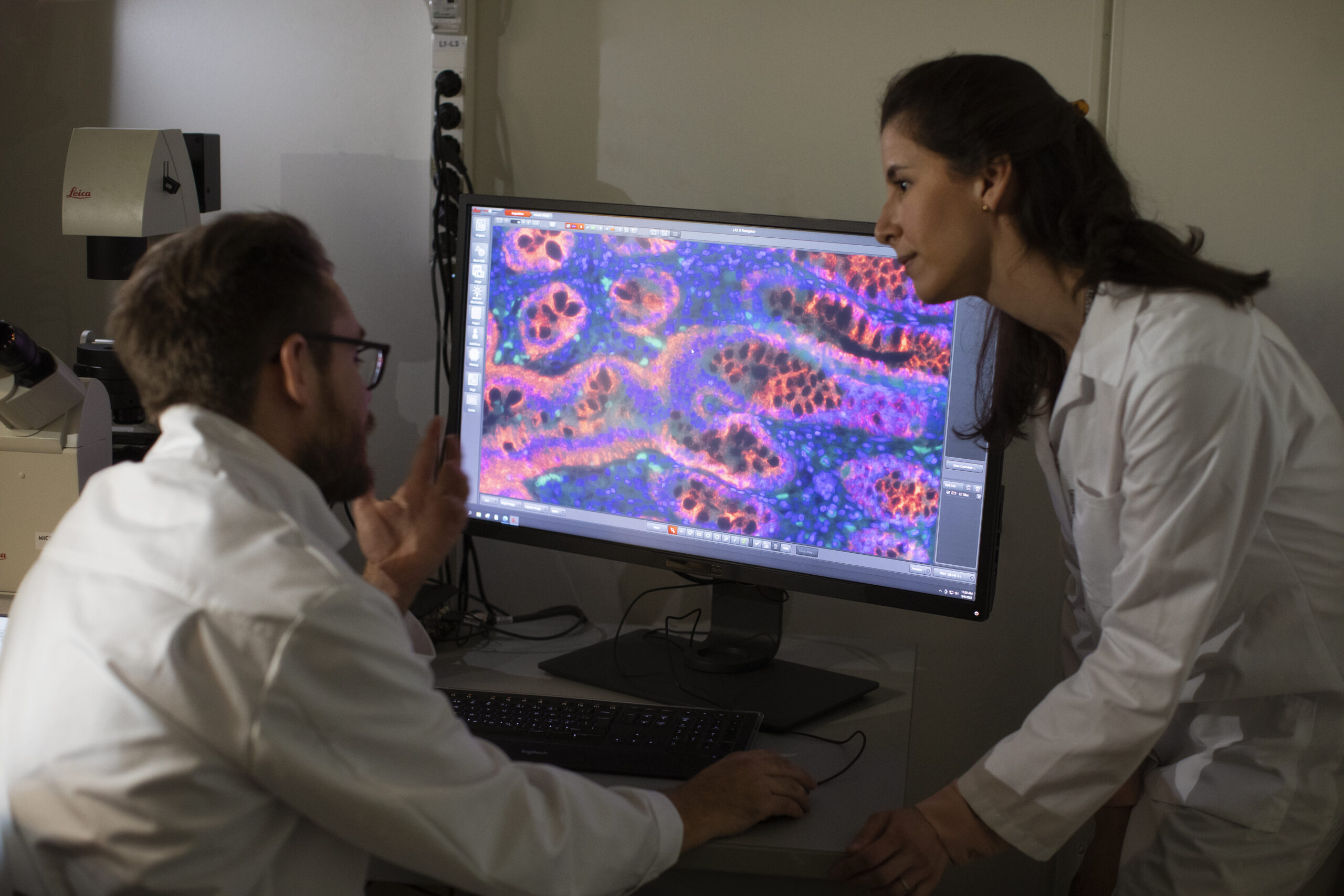
PD1/PD-L1 interaction detected with NaveniFlex Tissue